Home » UnlabelledDiagram Of The Muscles In The Forearm - Diagram Tendons In Arm Diagram Full Version Hd Quality Arm Diagram Ahadiagram Innesti Grafting It - The accompanying muscle diagram reveals the muscles' positions beneath the surface.
Jumat, 25 Juni 2021
Diagram Of The Muscles In The Forearm - Diagram Tendons In Arm Diagram Full Version Hd Quality Arm Diagram Ahadiagram Innesti Grafting It - The accompanying muscle diagram reveals the muscles' positions beneath the surface.
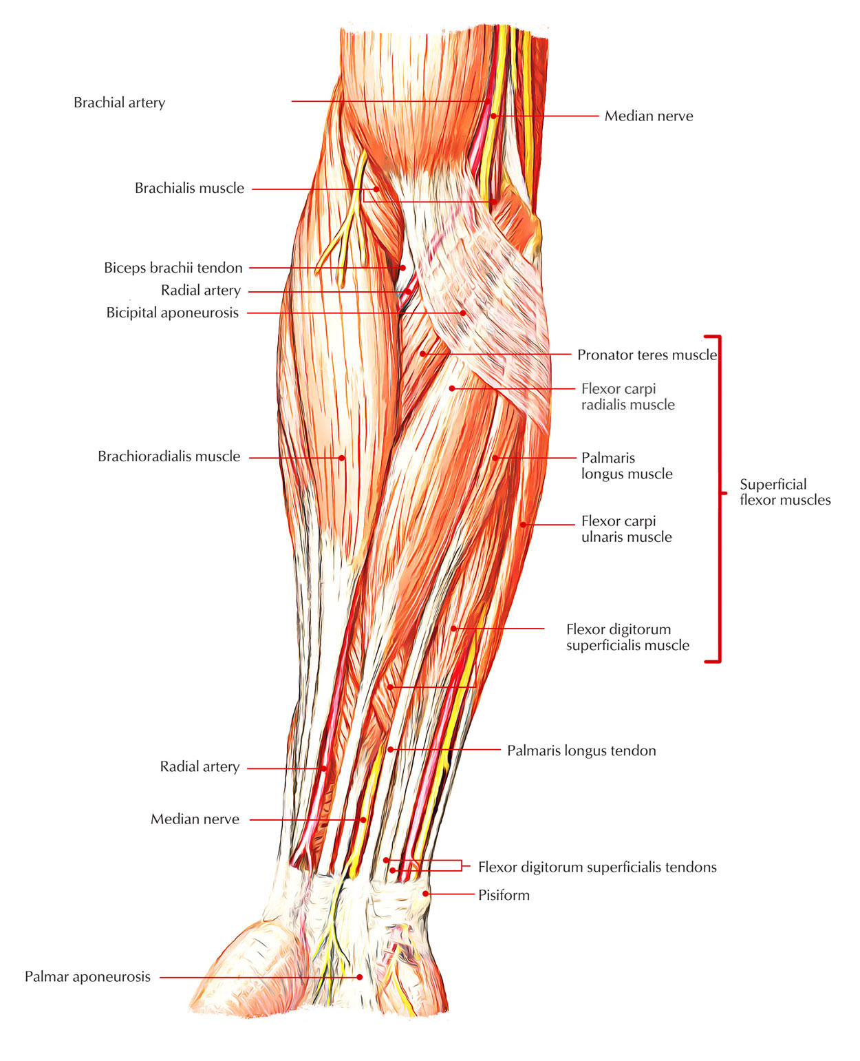
Diagram Of The Muscles In The Forearm - Diagram Tendons In Arm Diagram Full Version Hd Quality Arm Diagram Ahadiagram Innesti Grafting It - The accompanying muscle diagram reveals the muscles' positions beneath the surface.. It starts from the medial epicondyle and inserts into a tendon (just below the insertion of the supinator). The antibrachial or forearm muscles may be divided into a volar and a dorsal group. Pronator teres pronates the forearm, turning the hand posteriorly. The forearm is the region of the upper limb between the elbow and the wrist. Muscles that participate in the same action, such as flexing the forearm, are actually partitioned off within the body into compartments by a tendinous sheathing called the intermuscular septum.
Pronator teres pronates the forearm, turning the hand posteriorly. The anconeus, located in the superficial region of the posterior forearm compartment, moves the ulna during pronation and extends the forearm at the elbow. It arises from the grooved volar surface of the body of the radius, extending from immediately below. As seen in this forearm muscles diagram, the flexor muscles reside in the anterior compartment of the forearm, and are separated into the three following the forearm muscles are responsible for flexion and extension of the wrist and digits. It is a functionally important muscle that contains two heads.
Easy Notes On Muscles Of The Anterior Or Front Of The Forearm Earth S Lab from www.earthslab.com The muscles of the forearm are about equally divided between those that cause movements at the wrist and those that move the fingers and thumb. The forearm is the region of the upper limb between the elbow and the wrist. A deep layer , intermediate layer and superficial layer. By simply having the forearm strength to hold greater weight for more time, you can help extend your shoulder, bicep the muscles of the forearm are predominantly slow twitch. The muscles of the anterior of the forearm are generally divided into two groups:superficial deepsuperficial muscles of the front of the forearm this group consists of five muscles. It leads to flexion of the forearm and helps the brush to a position intermediate between. 4, attachment… the muscles of the back forearm. Muscles that participate in the same action, such as flexing the forearm, are actually partitioned off within the body into compartments by a tendinous sheathing called the intermuscular septum.
It is a functionally important muscle that contains two heads.
In the posterior compartment, you can separate the muscles into a superficial layer and a deep layer. In the anterior compartment, they are split into three categories: Diagram the movements of the humerus muscles that act on the forearm. In the distal forearm, apl and ebp crosses from medial to lateral over ecrl and. Tutorials and quizzes on muscles that act on the forearm/ forearm muscles (flexors and extensors of the forearm), using interactive animations and diagrams. Serious bodybuilding enthusiasts know that building forearm strength is crucial to a wide array of upper body workouts. Start studying muscles of the forearm. Learn vocabulary, terms and more with flashcards, games and other study tools. The term forearm is used in anatomy to distinguish it from the arm. Pronator teres pronates the forearm, turning the hand posteriorly. Longus, brevis, longus, brevis (longus is lateral to brevis). Related posts of muscles of the arm and forearm diagram. Inflammation of this region caused by repetitive.
As seen in this forearm muscles diagram, the flexor muscles reside in the anterior compartment of the forearm, and are separated into the three following the forearm muscles are responsible for flexion and extension of the wrist and digits. The term forearm is used in anatomy to distinguish it from the arm. Diagram the movements of the humerus muscles that act on the forearm. The superficial extensors of the forearm are the brachioradialis, extensor carpi radialis longus, anconeus, extensor carpi radialis brevis, extensor carpi ulnaris, extensor digitorum and extensor digiti minimi. The human muscular system is complex and has many functions in the body.
Rahul S Medical Images Medical Images Upper Extremities Forearm Muscles 2 Jpg from rahulgladwin.com The term forearm is used in anatomy to distinguish it from the arm. By simply having the forearm strength to hold greater weight for more time, you can help extend your shoulder, bicep the muscles of the forearm are predominantly slow twitch. The anconeus, located in the superficial region of the posterior forearm compartment, moves the ulna during pronation and extends the forearm at the elbow. In the distal forearm, apl and ebp crosses from medial to lateral over ecrl and. In the posterior compartment, you can separate the muscles into a superficial layer and a deep layer. Muscle anatomy diagram 12 photos of the muscle anatomy diagram canine muscle anatomy diagram, dog muscle anatomy diagram, lower leg muscle anatomy diagram, muscle anatomy of human back, tricep muscle. It is the weakest type of muscle but has an essential role in moving food along the digestive tract and. This is the most medial of the superficial flexor muscles in the forearm.
Remembering the action of each one can be quite difficult.
The forearm is divided into two compartments, which are separated by the radius and ulna and the interosseous membrane running between them. Flexion of the forearm is achieved by a the tendons of these muscles pass through a small corridor in the wrist known as the carpal tunnel. The human muscular system is complex and has many functions in the body. This layer contains only one muscle, the flexor digitorum. A deep layer , intermediate layer and superficial layer. Muscle anatomy diagram 12 photos of the muscle anatomy diagram canine muscle anatomy diagram, dog muscle anatomy diagram, lower leg muscle anatomy diagram, muscle anatomy of human back, tricep muscle. Try labeling diagrams and worksheets as additional learning aids. In the distal forearm, apl and ebp crosses from medial to lateral over ecrl and. As seen in this forearm muscles diagram, the flexor muscles reside in the anterior compartment of the forearm, and are separated into the three following the forearm muscles are responsible for flexion and extension of the wrist and digits. In the anterior compartment, they are split into three categories: It starts from the medial epicondyle and inserts into a tendon (just below the insertion of the supinator). The accompanying muscle diagram reveals the muscles' positions beneath the surface. Diagram of the muscles of the arm in action.
This layer contains only one muscle, the flexor digitorum. The muscles of the upper arm are responsible for the flexion and extension of the forearm at the elbow joint. Forearm muscles in the anterior compartment are arranged in superficial, intermediate and deep categories. Start studying muscles of the forearm. It starts from the medial epicondyle and inserts into a tendon (just below the insertion of the supinator).
Forearm Wikipedia from upload.wikimedia.org The human muscular system is complex and has many functions in the body. Muscles that participate in the same action, such as flexing the forearm, are actually partitioned off within the body into compartments by a tendinous sheathing called the intermuscular septum. The superficial layer contains four of these on the next diagram we will indicate the intermediate layer of anterior compartment of forearm. Pronator teres pronates the forearm, turning the hand posteriorly. There are many muscles in the forearm. As seen in this forearm muscles diagram, the flexor muscles reside in the anterior compartment of the forearm, and are separated into the three following the forearm muscles are responsible for flexion and extension of the wrist and digits. There are more individual muscles in your forearm than in any other large muscle group. The superficial extensors of the forearm are the brachioradialis, extensor carpi radialis longus, anconeus, extensor carpi radialis brevis, extensor carpi ulnaris, extensor digitorum and extensor digiti minimi.
The forearm is divided into two compartments, which are separated by the radius and ulna and the interosseous membrane running between them.
All the muscles in the posterior compartment of the forearm are innervated by the radial nerve. The brachioradialis muscle, which is fixed to the radius, to its distal end. Smooth muscle lines the inside of blood vessels and organs, such as the stomach, and is also known as visceral muscle. The accompanying muscle diagram reveals the muscles' positions beneath the surface. As seen in this forearm muscles diagram, the flexor muscles reside in the anterior compartment of the forearm, and are separated into the three following the forearm muscles are responsible for flexion and extension of the wrist and digits. The forearm is the region of the upper limb between the elbow and the wrist. The superficial extensors of the forearm are the brachioradialis, extensor carpi radialis longus, anconeus, extensor carpi radialis brevis, extensor carpi ulnaris, extensor digitorum and extensor digiti minimi. The term forearm is used in anatomy to distinguish it from the arm. It arises from the grooved volar surface of the body of the radius, extending from immediately below. Inflammation of this region caused by repetitive. Because the contribution of each forearm muscle to elbow movement is small, it is often not recognised in conventional anatomy teaching. The flexor pollicis longus is situated on the radial side of the forearm, lying in the same plane as the preceding. In the anterior compartment, they are split into three categories:
