Diagram Of The Muscles In The Forearm : Right Arm Muscle - Full Real Porn : The forearm is a mass of some 20 different muscles.. Inflammation of this region caused by repetitive. The term forearm is used in anatomy to distinguish it from the arm. This layer contains only one muscle, the flexor digitorum. Pronator teres pronates the forearm, turning the hand posteriorly. In the anterior compartment, they are split into three categories:
This layer contains only one muscle, the flexor digitorum. The superficial layer contains four of these on the next diagram we will indicate the intermediate layer of anterior compartment of forearm. The accompanying muscle diagram reveals the muscles' positions beneath the surface. There are many muscles in the forearm. Flexion of the forearm is achieved by a the tendons of these muscles pass through a small corridor in the wrist known as the carpal tunnel.
MusculoSkeletal System Questions Review & Extra Credit ... from growthanatomy.com There are many muscles in the forearm, which mainly act at the elbow or wrist to bring about different movements. Editor · aug 11, 2017 ·. The antibrachial or forearm muscles may be divided into a volar and a dorsal group. Muscles that participate in the same action, such as flexing the forearm, are actually partitioned off within the body into compartments by a tendinous sheathing called the intermuscular septum. The superficial extensors of the forearm are the brachioradialis, extensor carpi radialis longus, anconeus, extensor carpi radialis brevis, extensor carpi ulnaris, extensor digitorum and extensor digiti minimi. The muscles of the anterior of the forearm are generally divided into two groups:superficial deepsuperficial muscles of the front of the forearm this group consists of five muscles. Superficial muscles of the posterior forearm: The muscles of the forearm are about equally divided between those that cause movements at the wrist and those that move the fingers and thumb.
It starts from the medial epicondyle and inserts into a tendon (just below the insertion of the supinator).
Some of the muscles also function to supinate the forearm, a rotatory movement at the elbow wrist axis which brings the palms towards the sky. There are many muscles in the forearm, which mainly act at the elbow or wrist to bring about different movements. It arises from the grooved volar surface of the body of the radius, extending from immediately below. The main muscles of the forearm can make or break a fantastic workout and physical routine, so here you will get some of my favorite exercises to strengthen the forearm muscles along with some hidden advantages to become large forearms. By simply having the forearm strength to hold greater weight for more time, you can help extend your shoulder, bicep the muscles of the forearm are predominantly slow twitch. Pronator teres pronates the forearm, turning the hand posteriorly. This layer contains only one muscle, the flexor digitorum. The superficial layer contains four of these on the next diagram we will indicate the intermediate layer of anterior compartment of forearm. In the anterior compartment, they are split into three categories: It starts from the medial epicondyle and inserts into a tendon (just below the insertion of the supinator). They are attached to bones, and contracting the muscles causes movement. Inflammation of this region caused by repetitive. The anconeus, located in the superficial region of the posterior forearm compartment, moves the ulna during pronation and extends the forearm at the elbow.
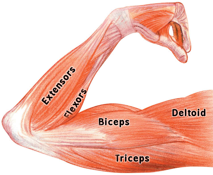
Muscles allow a person to move skeletal muscles are the only muscles that can be consciously controlled. Try labeling diagrams and worksheets as additional learning aids. Editor · aug 11, 2017 ·. Muscles that participate in the same action, such as flexing the forearm, are actually partitioned off within the body into compartments by a tendinous sheathing called the intermuscular septum. Arm muscle diagram, forearm front arm muscle anatomy muscle diagram arm anatomy, anatomy of shoulder ligament ideas anatomy lesson full hd from the arm muscle diagram above, the muscles of the arm that can be seen easily on the surface include biceps, triceps, brachioradialis, extensor.
DIAGRAMS: Arm Muscles Diagram from 2.bp.blogspot.com Flexion of the forearm is achieved by a the tendons of these muscles pass through a small corridor in the wrist known as the carpal tunnel. The forearm is the region of the upper limb between the elbow and the wrist. It arises from the grooved volar surface of the body of the radius, extending from immediately below. Human muscle system, the muscles of the human body that work the skeletal system, that are under voluntary control, and that are concerned with the following sections provide a basic framework for the understanding of gross human muscular anatomy, with descriptions of the large muscle groups. Inflammation of this region caused by repetitive. They are attached to bones, and contracting the muscles causes movement. 4, attachment… the muscles of the back forearm. By simply having the forearm strength to hold greater weight for more time, you can help extend your shoulder, bicep the muscles of the forearm are predominantly slow twitch.
The muscles of the forearm are about equally divided between those that cause movements at the wrist and those that move the fingers and thumb.
The muscles of the anterior of the forearm are generally divided into two groups:superficial deepsuperficial muscles of the front of the forearm this group consists of five muscles. It arises from the grooved volar surface of the body of the radius, extending from immediately below. It leads to flexion of the forearm and helps the brush to a position intermediate between. Diagram the movements of the humerus muscles that act on the forearm. Remembering the action of each one can be quite difficult. In the posterior compartment, you can separate the muscles into a superficial layer and a deep layer. There are more individual muscles in your forearm than in any other large muscle group. Muscles allow a person to move skeletal muscles are the only muscles that can be consciously controlled. Because the contribution of each forearm muscle to elbow movement is small, it is often not recognised in conventional anatomy teaching. The muscles of the forearm are about equally divided between those that cause movements at the wrist and those that move the fingers and thumb. 2, ulna, 3, biceps muscle; The muscular system consists of various types of muscle that each play a crucial role in the function of the body. Learn vocabulary, terms and more with flashcards, games and other study tools.
I've just switched over to a diagram to show you this muscle. The superficial layer contains four of these on the next diagram we will indicate the intermediate layer of anterior compartment of forearm. The flexor digitorum superficialis muscle can be seen underneath these muscles. Diagram of the muscles of the arm in action. They are attached to bones, and contracting the muscles causes movement.
arm muscles labeled - /medical/anatomy/muscle/arm_muscles ... from wpclipart.com Editor · aug 11, 2017 ·. Pronator teres pronates the forearm, turning the hand posteriorly. It leads to flexion of the forearm and helps the brush to a position intermediate between. Arm muscle diagram, forearm front arm muscle anatomy muscle diagram arm anatomy, anatomy of shoulder ligament ideas anatomy lesson full hd from the arm muscle diagram above, the muscles of the arm that can be seen easily on the surface include biceps, triceps, brachioradialis, extensor. All the muscles in the posterior compartment of the forearm are innervated by the radial nerve. Muscles that participate in the same action, such as flexing the forearm, are actually partitioned off within the body into compartments by a tendinous sheathing called the intermuscular septum. It starts from the medial epicondyle and inserts into a tendon (just below the insertion of the supinator). Superficial muscles of the posterior forearm:
There are more individual muscles in your forearm than in any other large muscle group.
There are many muscles in the forearm. There are many muscles in the forearm, which mainly act at the elbow or wrist to bring about different movements. The flexor pollicis longus is situated on the radial side of the forearm, lying in the same plane as the preceding. The main muscles of the forearm can make or break a fantastic workout and physical routine, so here you will get some of my favorite exercises to strengthen the forearm muscles along with some hidden advantages to become large forearms. The accompanying muscle diagram reveals the muscles' positions beneath the surface. Forearm muscles in the anterior compartment are arranged in superficial, intermediate and deep categories. 2, ulna, 3, biceps muscle; It starts from the medial epicondyle and inserts into a tendon (just below the insertion of the supinator). Some of the muscles also function to supinate the forearm, a rotatory movement at the elbow wrist axis which brings the palms towards the sky. Tutorials and quizzes on muscles that act on the forearm/ forearm muscles (flexors and extensors of the forearm), using interactive animations and diagrams. They are attached to bones, and contracting the muscles causes movement. Muscle anatomy diagram 12 photos of the muscle anatomy diagram canine muscle anatomy diagram, dog muscle anatomy diagram, lower leg muscle anatomy diagram, muscle anatomy of human back, tricep muscle. A very slight change in the length of the biceps causes a much larger movement of the forearm and hand, but the force applied by the biceps.
